
Eye Pain Causes

What Causes Eye Pain ?
The human eye is a beautifully complex structure. An entire lobe of the brain is dedicated to the processing of information transmitted from the eye. Furthermore, that information is “networked” to various locations throughout the brain. What could it mean when this miraculous organ signals disorder with pain? This post will be dedicated to explaining eye pain causes.
From the earliest written accounts by ancient cultures, mystical significance has been often attributed to the eye. Indeed, the saying, “the eye is the window to the soul” illustrates how mankind views this magical organ system.
Vision itself is not just the transmission of a detectable image but is also a complex process that ties in all that it means to be human. Collectively this is called visual perception.
Visual perception is very individualized. We do not all perceive the same way, though we may all be looking at the same object. Magicians use this processing to their advantage as they manipulate visual cueing by sleight of hand.
While the process of vision is a miracle in perception, there are limitations to it. Spectrum of light, speed of movement, distractibility of the observer, and emotional temperament all effect what is seen. Indeed, the professional baseball player never actually sees the fast ball come over the plate. The image is lost some 30 feet before the ball crosses the plate.

How is the ball then hit, you may ask? The expert batter estimates where the ball will cross home plate as he swings at an invisible ball. Given the contact area between the ball and the bat is a few millimeters in width and length, the assertion that hitting a baseball fastball is the most difficult action in all of sports is well deserved.
Pain of the eye is never normal. Though pain can be expected in other organ systems through exercise or disuse, the eye is a mostly “solid state” organ. Its visual receptor apparatus has no moving parts (the retina) and so can withstand long periods of use without exhaustion.
The remainder of this post will be to help you discern what may be causing your or another’s eye pain.
This is important
IN MY OPINION, ALL PERISTENT EYE PAIN REQUIRES THE IMMEDIATE EVALUATION BY A LICENSED, QUALIFIED MEDICAL PRACTITIONER.
A BRIEF REVIEW OF EYE ANATOMY AND PHYSIOLOGY
The anatomy of the eye can be divided into two sets of structures…those that guide the reflected light from a source image are different from the structures that transduce, translate, and transmit the image.
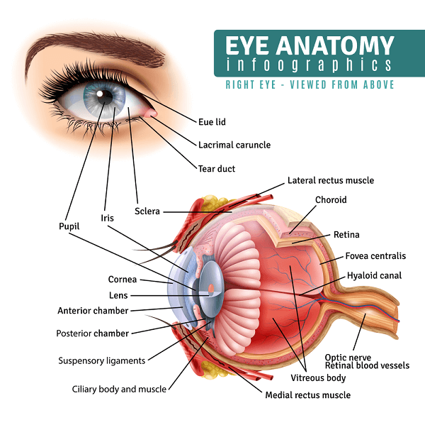
The image guiding structures are:
- Cornea: the clear “dime sized” structure that allows light to enter the eye.
- Iris: the colored circular muscle that opens and closes like the shutter of a camera. It is the external feature that we admire most about the eye. The pupil is in the center.
- Sclera: the “white” of the eyeball. It makes up the globe that contains most of the structures of the eye. The symmetry (or lack of it) effects the images’ focus on the retina.
- Conjunctiva: a clear membrane that covers the sclera and underside of the eyelids.
- Lens: immediately behind the iris, it is a clear structure that helps focus the image on the retina.
- Eyelids: protective to the eye as well as mechanically spread the tears from the lateral margins of the eye surface to the middle of the eye surface.
- Extra-ocular eye muscles: they position the eye for best image capture.
- Orbit: the protective boney cup where the eye can rotate in a myriad of directions.
The image processing structures are:
- Retina: a delicate transducer (a transducer is any apparatus that changes one form of energy into another). Light energy is transduced into an electrical signal by the retina.
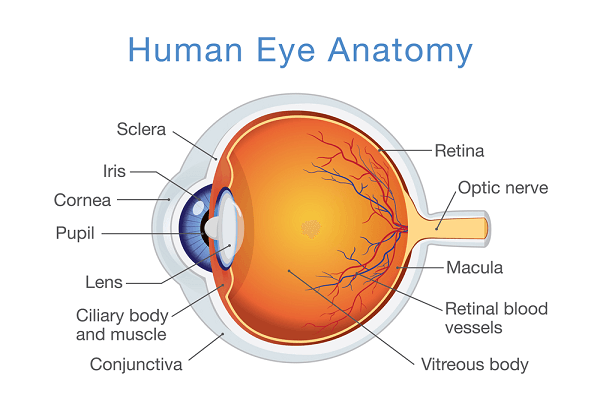
- Optic Nerve: transmits the electrical signal from the retina to the occipital lobe of the brain.
- Occipital Cortex of the Brain: without processing of a transmitted image there can be no visual perception. Though this is not anatomically part of the eye, vision cannot occur without it.
There are other anatomical parts to the eye which are beyond the scope of an article of this type.
Physiologically the retina is an extension of the brain. As a baby develops in utero, the retina extends from the primordial brain tissue to the areas where the eyes develop.
Exactly when in utero visual perception of the fetus actually occurs can be debated. However, at 18 weeks (the eyelids are still closed on the baby) bright light shined on the mother’s abdomen is perceived by the baby in her womb.
The light that enters the eye is focused onto the retina. The light energy activates the specialized cells in the back of the eye (in the retina) which is converted into electrical signaling. The signal travels along the optic nerve to the back of the brain (the occipital lobe of the brain) where it is multi-processed.
A very important aspect of visual perception is the “pruning” of the visual information. “Pruning” or trimming of the information actually begins in the retina. If all the visual information that entered the eye was actually processed the occurrence of visual perception would be much slower.
What would cause eye pain ?
here are many causes for eye pain. I will cover the top 3 causes:
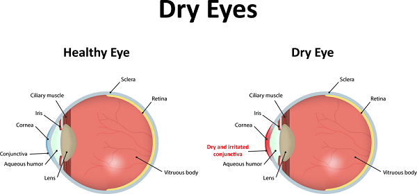
1) Eye Dryness:
There is a minimal amount of tear production that must occur for the eye surface to remain lubricated. If the production is reduced below this minimum then the feeling of “grittiness” occurs when the eyes are blinked. The lubrication is essential for the surface of the eye, particularly the cornea, to not dry out. This type of eye pain is not incapacitating and is generally not associated with loss of vision.
2) A Foreign Body in the eye:
Any debris from the environment can become lodged or caught on the surface of the eye. The pain is usually quite severe and sudden. Excessive tearing usually occurs in response. Curiously, due to the neural connections of the eyes, a foreign body in one eye will actually make both eyes tear excessively. There is not usually a loss of vision (though the vision may be distorted by the tearing).
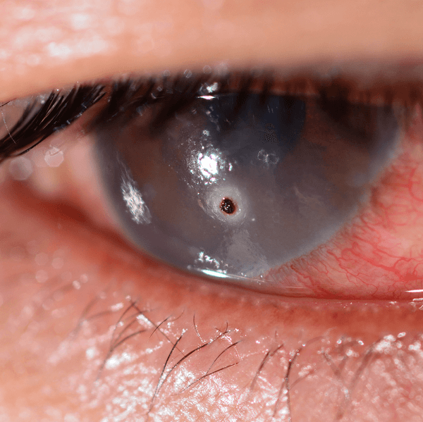
3) Conjunctivitis:
This is a condition where the thin membrane covering the outside of the eye (and inner eyelids) becomes inflamed or reddened. The cause can be a virus (the most common cause), a bacteria, or even allergic. The white of the eye appears pink and there is usually a moderate amount of discomfort (not excruciating). There is usually not a loss of vision.

HOW DO YOU DIAGNOSE EYE PAIN AND TREAT IT?
Important Note
A LOSS OF VISION (IT DOES NOT HAVE TO BE COMPLETE) IS AN EYE EMERGENCY AND REQUIRES IMMEDIATE EVALUATION BY A LICENSED MEDICAL PRACTITIONER.
- Eye dryness is diagnosed by a thorough history and physical examination of the eye. A thorough examination of the eye should always include an exam of the eye surface (with eversion of the eyelids to inspect the undersurface of the lids). Furthermore, an opthalmoscopic exam (either with a hand held scope or a Slit-lamp) is required.
Sufficient tear production is measured by a simple test called a Schirmer’s test (click here to link to how it is done). Essentially the test places a thin strip of absorbing paper in the lower lid recess and measures how much tear fluid is absorbed.
Treatment is essentially treatment of the underlying cause and tear replacement (eye drops). In some cases medications that will boost tear production can be given. An Ophthalmologist (an eye doctor) will be the person to diagnose and prescribe the appropriate medication.
- A foreign body on the eye surface must be removed. Simple removals are usually done in the emergency room by a trained staff person. More complex removals (such as a foreign body that may have pierced through the eye) are handled by an Ophthalmologist. An emergency room physician will know when to refer you to an Ophthalmologist.
- A simple conjunctivitis is usually treated by a primary care practitioner. Although most infections are self-limiting, antibiotic eye drops are the normal course of treatment. More severe eye infections, infections that don’t get better with antibiotic eye drops, or infections with a loss of vision are usually referred to an Ophthalmologist.
This is Important
ALL EYE PAIN DISORDERS REQUIRE A THOROUGH HISTORY AND PHYSICAL. AN EYE EXAMINATION ALWAYS INCLUDES A SURFACE EVALUATION (WITH EYELID EVERSION) AS WELL AS AN INTERNAL EYE EXAM (WITH AN OPTHALMOSCOPE AND/OR A SLIT LAMP EXAM).
THERE IS HOPE
I have reviewed 3 eye pain causes (there are many more). Any pain in the special sense organs requires professional evaluation. The threshold for referral should be low in eye disorders. Unless the cause is obvious and easily treated, seek the prompt evaluation by an Ophthalmologist.
I hope this post has been informative and helpful. Please leave me a comment or question below. I would love to hear from you.
Wishing you good health and peace,
Facebook
Google+
Twitter
Pinterest
Reddit
StumbleUpon